views
Drawing the Model
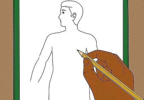
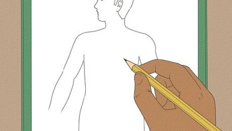
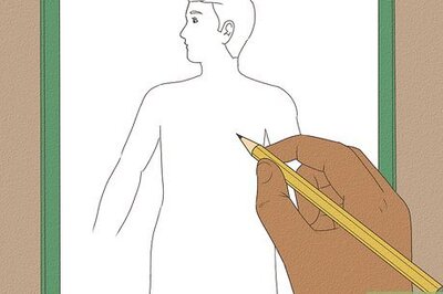
Start by drawing an outline of a person. You only need to draw the head and torso. Make sure you are using a pencil rather than a pen so that you can erase if necessary. This outline should take up most of the space on your paper. Draw the head in proportion to the body as it would be on a human. It doesn’t have to be a very involved or elaborate outline, a simple circle for the head and somewhat rectangular torso will do. This is just to provide a frame of reference for your digestive model. Draw the head as a profile rather than straight on to make it easier to show the digestive organs in the head. If you wish, feel free to be creative and embellish this outline a bit. You can draw eyes and a nose and ears and hair. You could even give your person a name if you feel like it! Just don’t draw over the torso or you will obscure your model.
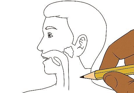
Add the mouth, teeth, and tongue. You can draw the mouth open as a sideways “v” and add a small curve on the bottom for a tongue and some small squares for teeth on the top. Your first step of digestion is now complete! Digestion begins in the mouth with ingestion. Salivary glands release saliva containing digestive enzymes to begin breaking down food while you chew. The tongue helps the food to move back in your throat creating a bolus while the teeth break it down in a process known as mastication.
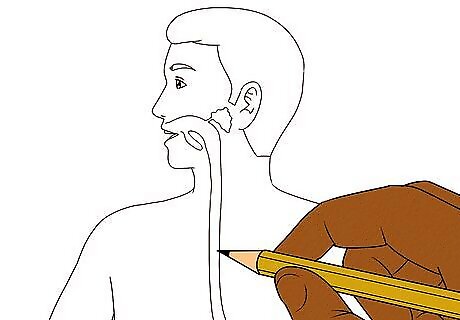
Draw the esophagus. At the end of the mouth, draw a small tube that extends straight down into the center of your model’s torso. It should be fairly narrow, about 1/5 the width of your model's neck. The food moves from the mouth into the esophagus, which carries it down into the stomach. The esophagus is made of smooth muscle that relaxes and contracts to move your food down with a wavelike motion, called peristalsis. If you wish to make your model more advanced, you can include the pharynx. The pharynx is located behind the mouth and moves food into the esophagus. You can feel it when you swallow. In your model, you can simply draw a small diagonal line towards the top of your esophagus and the part above the line can be the pharynx and the part below can be the esophagus. It you wish to make your model more advanced, you can also include the epiglottis. The epiglottis is a small flap just below the pharynx that directs food into the esophagus. If you drew a pharynx, the diagonal line can be the epiglottis.
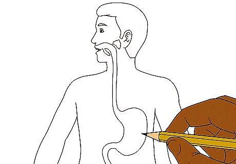
Include the stomach. At the bottom of your esophagus, draw a small balloon-like organ that is the stomach. It should take up about a third of the width of the torso and be slightly towards the right. Include a small tube going up into the esophagus and a diagonal line between the esophagus and stomach to indicate the muscular valve called the lower esophageal sphincter. Add a small tube going straight down on the left side of the stomach, which will lead to the small intestine. The stomach helps churn and digest food using gastric juices containing hydrochloric acid and pepsin to help break it down. The food remains in the stomach for about 3-4 hours. At this point it is no longer food, but has an oatmeal-like consistency and is called "chyme."
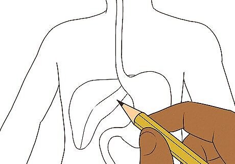
Add the liver. Draw it towards the left of the stomach and slightly above it. It should be about twice the size of the stomach and look a bit like an elongated triangle with rounded edges. The liver produces bile to help break down fats. While the food doesn’t enter the liver, it processes the nutrients that are absorbed from the small intestine.
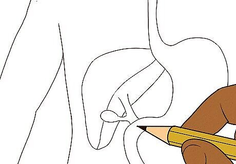
Draw the gallbladder. Attached to the liver, draw a small nodule called the gallbladder. Draw the gallbladder as a small oval towards the bottom of the liver. It should overlap the liver. To show that the gallbladder goes over the liver, draw the gallbladder with a thick line. The gallbladder stores the bile produced in the liver. It then mixes the bile with the food that passes by, breaking down fats.
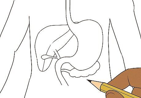
Sketch the pancreas. It should be a small carrot-shaped organ that goes behind the stomach. To show that the pancreas is behind the stomach, draw it with a dotted line. The pancreas releases digestive juices that aid in breaking down carbohydrates, protein, and fat as the food leaves the stomach. It also regulates blood sugar.
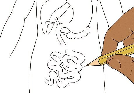
Include the small intestine. The small intestine is a large curving tube that you can draw as squiggly lines beneath the stomach in the center of the body, taking up about half of the width of the body. Draw it a couple of inches below the stomach because the large intestine will end up going on top of it. The end of the small intestine should be towards the bottom of your paper and towards the left side. Add a diagonal line to indicate the muscular valve, called the pyloric sphincter, between the duodenum of the small intestine and the stomach. The small intestine is about 18–22 feet (5–7 m) long! This is where the majority of digestion takes place. The small intestine contracts to move food through it with the help of bile secretions and pancreatic and intestinal juices, allowing it to absorb nutrients with villi. To make a more advanced model, you can distinguish the different parts of the small intestine. The duodenum is a small tube connecting the stomach to the jejunum. The jejunum is where the majority of digestion takes place and is the middle part of the small intestine. The ileum is the last region of the small intestine that connects to the large intestine.
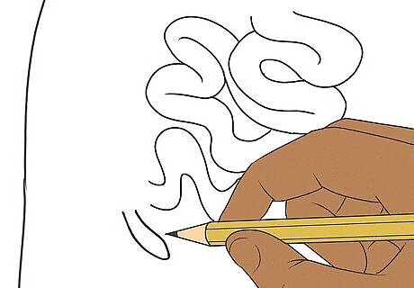
Add the appendix. It should be located at the end of the small intestine. Simply draw a small pouch at the end of the small intestine towards the left side of your paper. The appendix is a small pouch-like structure that is thought to have lost its purpose and become more of a liability than an aid in digestion. But it is part of the digestive system and sits where the small and large intestines connect.
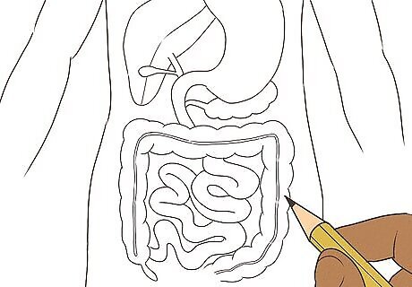
Sketch the large intestine. Extending from the end of the small intestine, right above the appendix, draw the large intestine. It should look like two squiggly tubes side by side and should go straight up on the left side of the small intestine, then go across the body below the stomach, and then go straight down towards the bottom of the body on the right. It should form the top three sides of a squiggly, tubelike- square. The passage of food is slower through the large intestine in order to allow for fermentation by bacteria and other microorganisms, called gut flora. The large intestine processes whatever cannot be used in digestion. It absorbs whatever it can, especially water, but then the rest will expelled as waste after about 12 hours. To make a more advanced model, distinguish between the different parts of the large intestine. The cecum is attached to the appendix and connects it to the ascending colon. The ascending colon goes straight upwards and connects to the transverse colon, which goes across the body. The transverse colon connects to the descending colon, which carries food down into the sigmoid colon, which goes directly to the rectum.
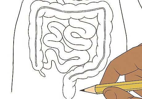
Draw the rectum and anus. At the end of the large intestine is the rectum. The rectum connects the large intestine to the anus. To draw these, simply draw a wide tube that is the rectum leading to a narrower tube that is the anus. This should go to the bottom of your sheet of paper. Congratulations, you have drawn a digestive model! The rectum stores feces until expulsion. The anus then expels waste.
Adding Finishing Touches
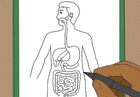
Outline your model. Now that your model is finished, take a black pen or marker and draw over it to create the final version. This is a nice finishing touch that will make it look more professional and will help it to really stand out. Simply trace over all of the pencil marks that you wish to keep.
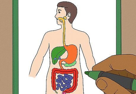
Color each organ a different color. This helps your model to stand out and also helps to distinguish between the organs. For some of the organs, simply by looking at the drawing, it can be hard to tell where one ends and another begins, but if you color code them, it will be clear. For overlapping organs, try using using lighter and darker shades of the same color. The areas where they overlap will be darker, and the areas where it is a single organ will be lighter.
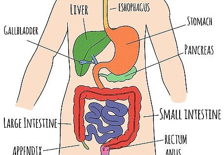
Label your model. Draw a thin line to each organ and write its name at the end of the line, outside of your model. This is great for reference so that you can study your model and learn which organ is which. If you prefer not to write the names of the organs on your model, you could instead make a key at the bottom or on another piece of paper where you draw a little square of each color and write the name of the organ next to it. This will allow you to quiz yourself on the names of the organs because they won’t be written right next to them. If you wish to make your model more advanced, you can differentiate the parts of the small intestine. Simply draw a small line towards the beginning of the small intestine to show where the duodenum turns into the jejunum and then draw a small line towards the end of the small intestine to show where the jejunum becomes the ileum. If you wish to make your model more advanced, you can also differentiate the parts of the large intestine. Simply draw a small line to divide each part. The cecum is attached to the appendix and connects it to the ascending colon. The ascending colon is the part that goes straight upwards. It connects to the transverse colon which goes across the body and is the largest part of the large intestine. The transverse colon connects to the descending colon, which carries food downward. Finally, there is the sigmoid colon which goes directly to the rectum.
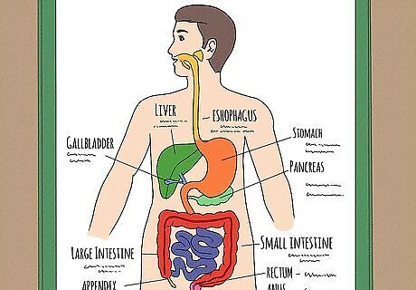
Write a brief description of each organ’s function. To learn even better, you can write a short description of each organ either with the labels or with your key. This will reinforce their functions and help your model to be very educational. It’s great to learn not just what the organs look like, but also their specific functions.




Comments
0 comment